FAMILY CHEYLETIDAE LEACH, 1815
Type genus: Cheyletus Latreille, 1776
Only representatives of 3 tribes are permanently associated with birds: Ornithocheyletiini and Metacheyletiini (parasites) and Cheletosomatini (predators and rarely parasites)
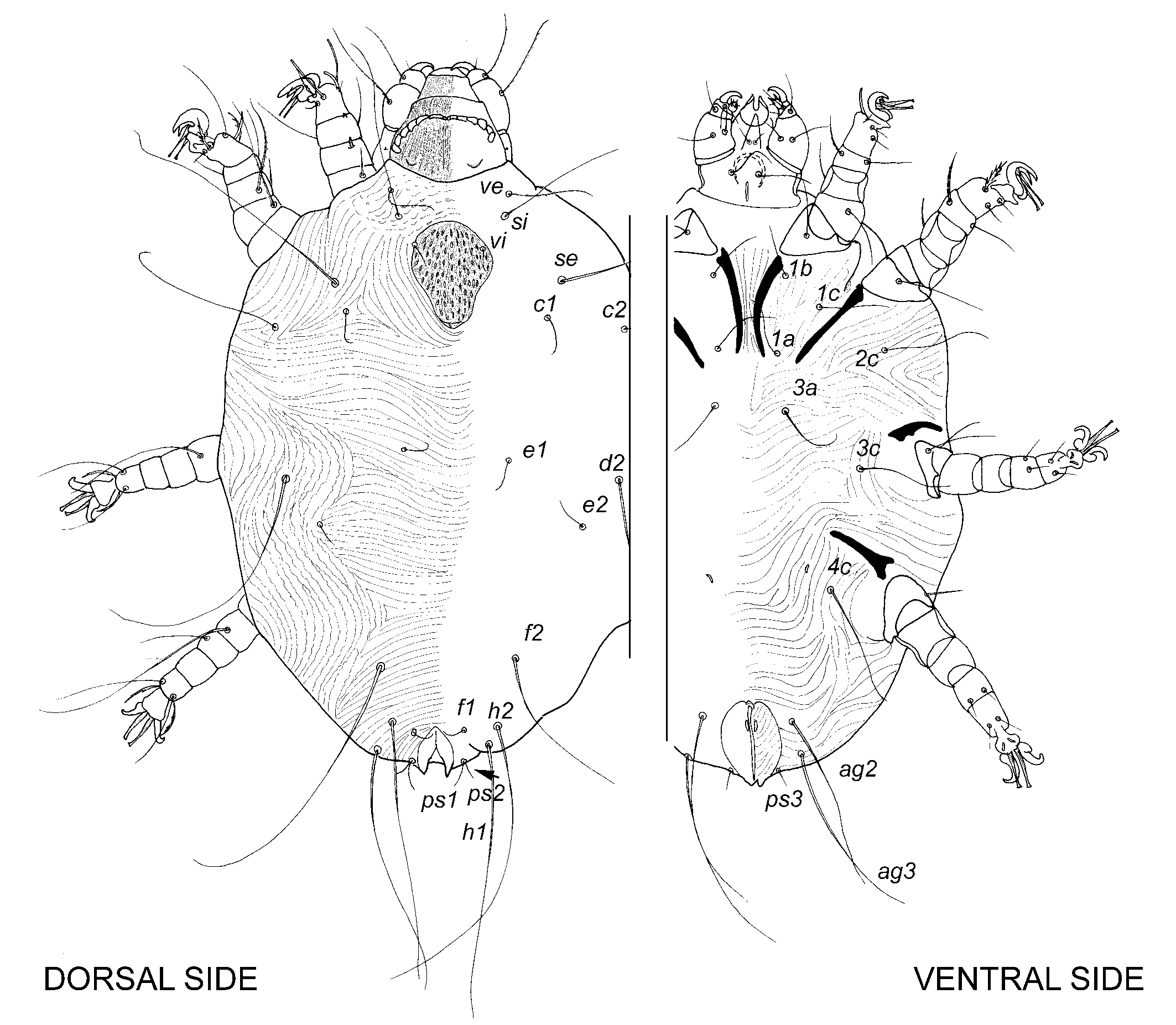
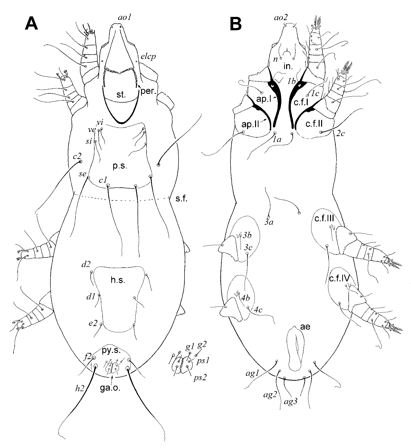
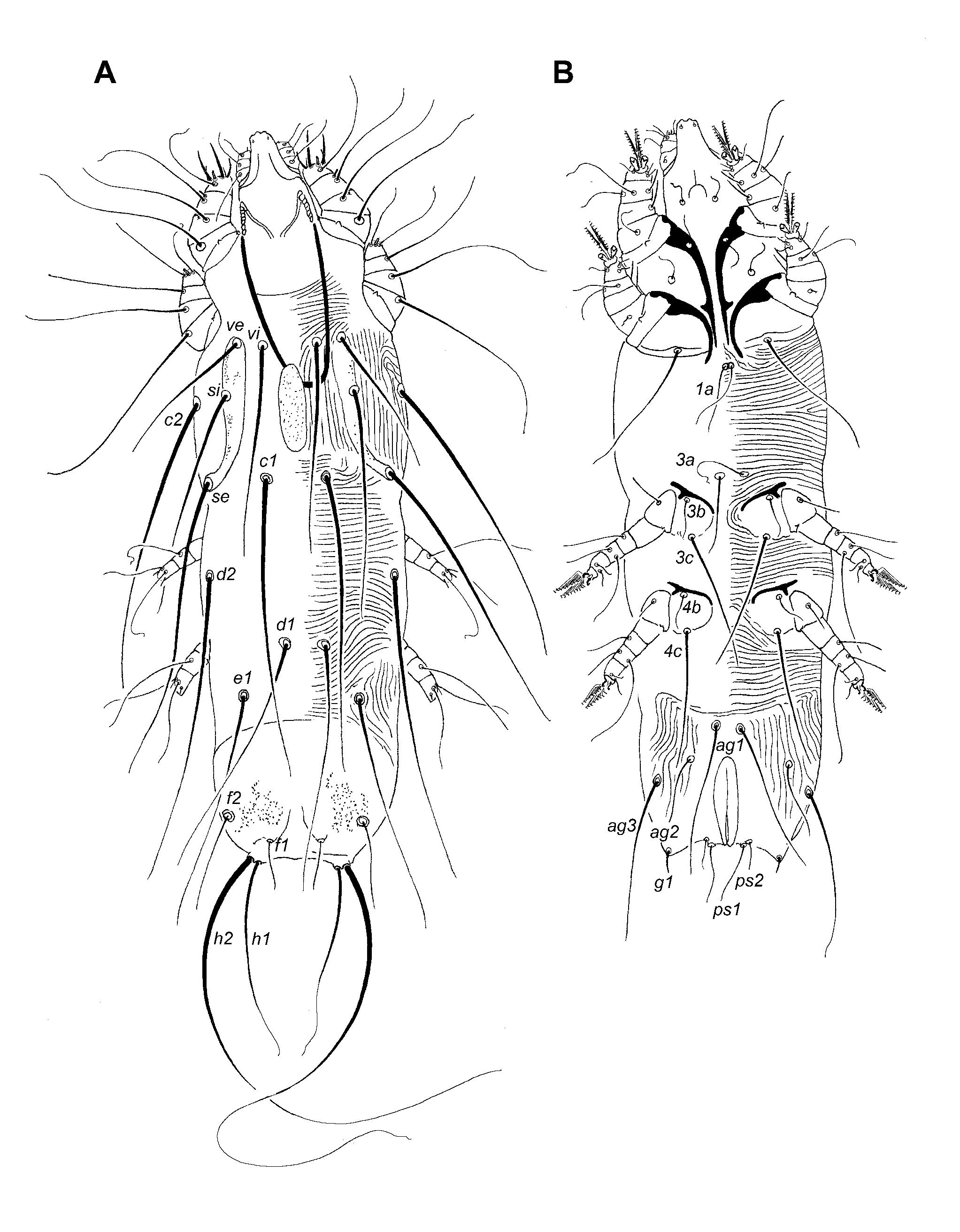
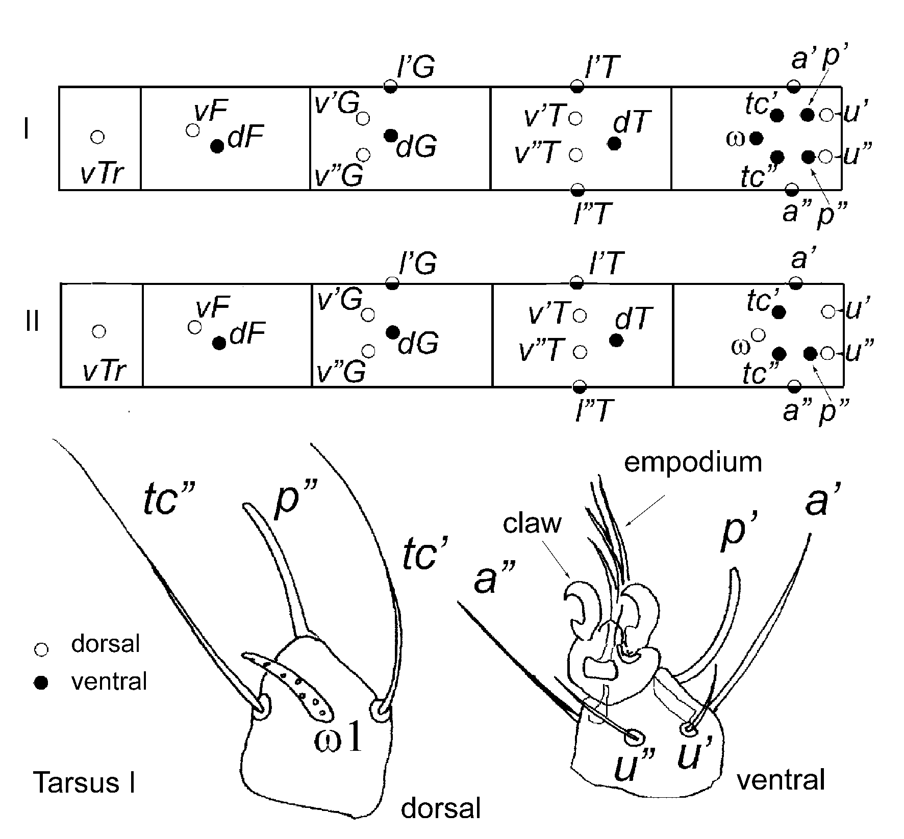
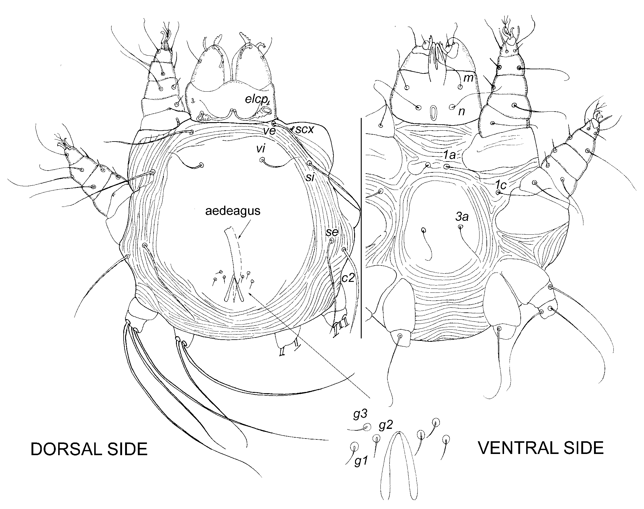
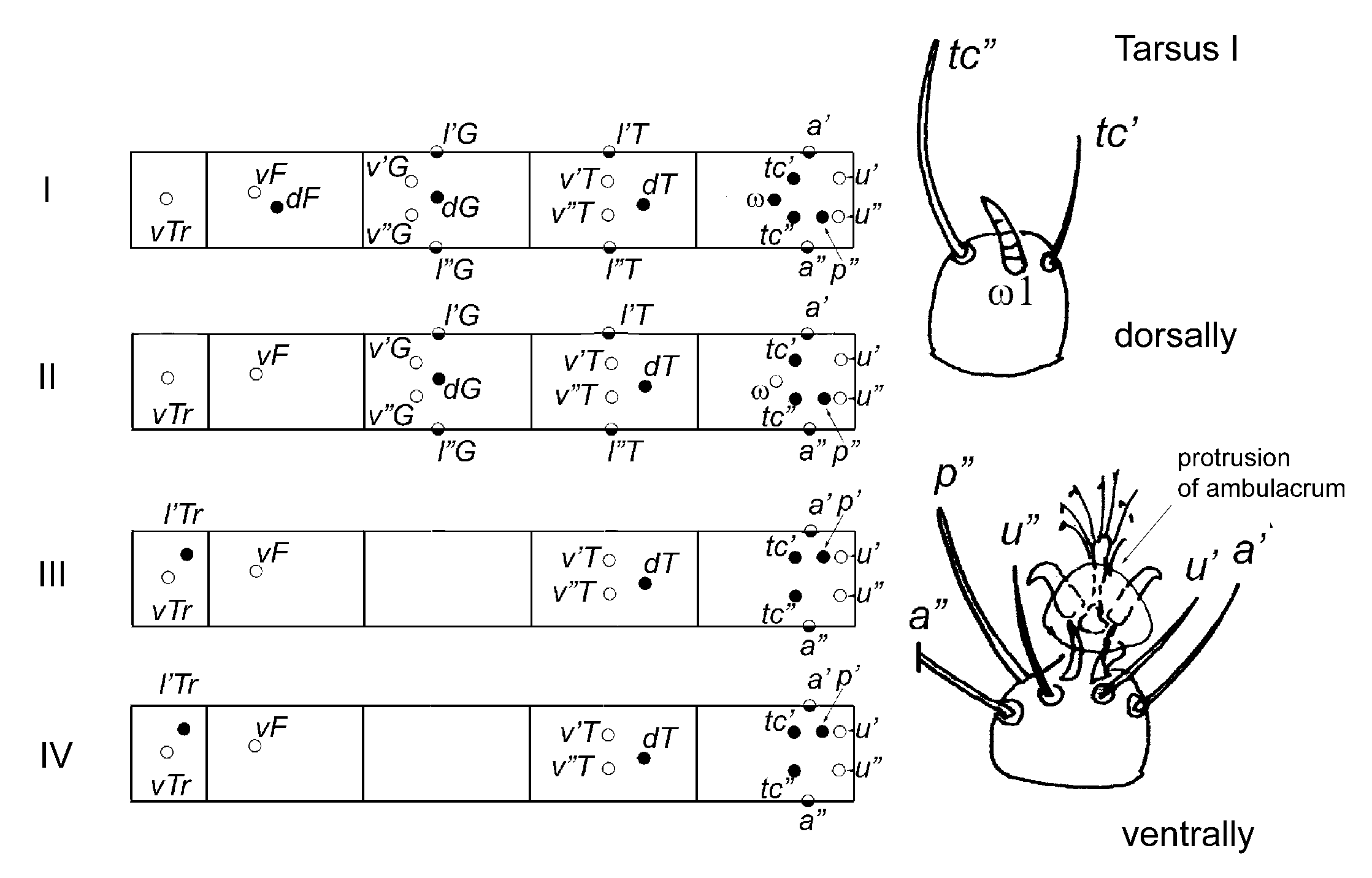
Description. Gnathosoma moderately or distinctly developed (1/5-1/3 of idisosomal length). Subcapitulum bearing setae elc p, n, ao1 and ao2. Peritremes situated on rostral part of stylophore, distinctly segmented, generally M-shaped. Distal part of hypostome fused with stylophore. Hypostomal lips present. Palps linear, consisting of 5 segments, with various projections in some parasitic forms. Palpal tibia with distinct claw. Palpal tarsus strongly reduced. Palpal setation: femur with setae d, v’, v”, genu - d, l”, tibia - d, l’, l”, and tarsus with eupathidia acm, sul, ul’, ul”, and solenidion ω. Eupathidia acm and sul” comb-like in most predaceous mites and smooth in most parasites. Idiosoma. Idiosoma generally flattened dorso-ventrally, rhomb-like or ovate in outline. Bases of trochanters inserted on ventral side of idiosoma. Eyes generally absent; one pair of lens-like eyes present on propodonotum in some free-living predators. Propodonotal shield present, distinctly developed. Hysteronotal and pygidial shields present or absent. Ventral side of idiosoma generally devoid of shields but in males of some predatory forms weakly sclerotized pseudosternal shield present. Opisthosoma moderately developed. In females, anal and genital-oviporal openings situated ventrally, close to posterior end of body or terminally, adnate and covered by pair of folds. In males, aedeagus tube-like or comma-like, pointed apically, genital and anal openings fused (or anal opening absent). In males of free-living forms, genital opening situated terminally or ventro terminally. In parasitic males, genital opening displaced dorsally, and in some bird parasites, it located in anterior part of idiosoma. Distance between coxal fields II and III distinctly shorter than idiosomal width (excluding tribe Bakini). In most taxa, 3 pairs of cupules present: im, ip, and ih. In some parasites, these cupules absent. Idiosomal setation (maximum set): scx, vi, ve, si, se, c1, c2, d1, d2, e1, e2, f1, f2, h1, h2, h3, ps1, ps2, ps3, ag1, ag2, ag3, g1, g2. Setae scx covered dorsally by extending lateral margins of propodonotum. In males, 3 pairs of aggenital setae, 2 pairs of genital setae, and 3 pairs of pseudoanal setae present. In some taxa idiosomal setae neotrichous and many neotrichous dorsal setae strongly modified. Legs. In most taxa, legs slender, consisting of 5 articulated segments, and their tarsi having weakly developed pretarsi bearing 2 smooth lateral claws and empodium with tenent hairs. Pretarsi separated from respective tarsi by dorsal knob. Leg length usually 60-70% of idiosomal length. In Metacheyletia, legs IV lost or primordial. In free-living forms, coxae distinctly bordered, and in some cases well sclerotized. In many parasitic mites, coxae represented exclusively by the coxal apodemes. Projections of leg segments present only in some parasitic species. Maximum set of leg setae (paired setae are in parentheses): tarsi I-IV — I, ft,(tc), a”, (p),(u), vs, ω1; II, (tc), (p),(u), vs, ω1; III, (tc), (p),(u), vs, ω1 (only in males); IV, (tc), (p),(u), vs, ω1 (only in males); tibiae I-IV – I, d, (l), (v),φ; II, d, l", (v), φ; III, d, l", (v), φ (only in males); IV, d, l", (v), φ (only in males); genua I-IV – I, d, l’, σ; II-IV, d, l’; femora I-IV – d, v; trochanters I-IV – I, II, IV, v; III, l, v; coxae I-IV – I, 1a, 1b, 1c; II, 2c; III, 3a, 3b, 3c; IV, 4a, 4b, 4c. Solenidion ω1II situated ventrally. Larva, proto- and tritonymphs present; deutonymph absent. Male moulting from protonymph.

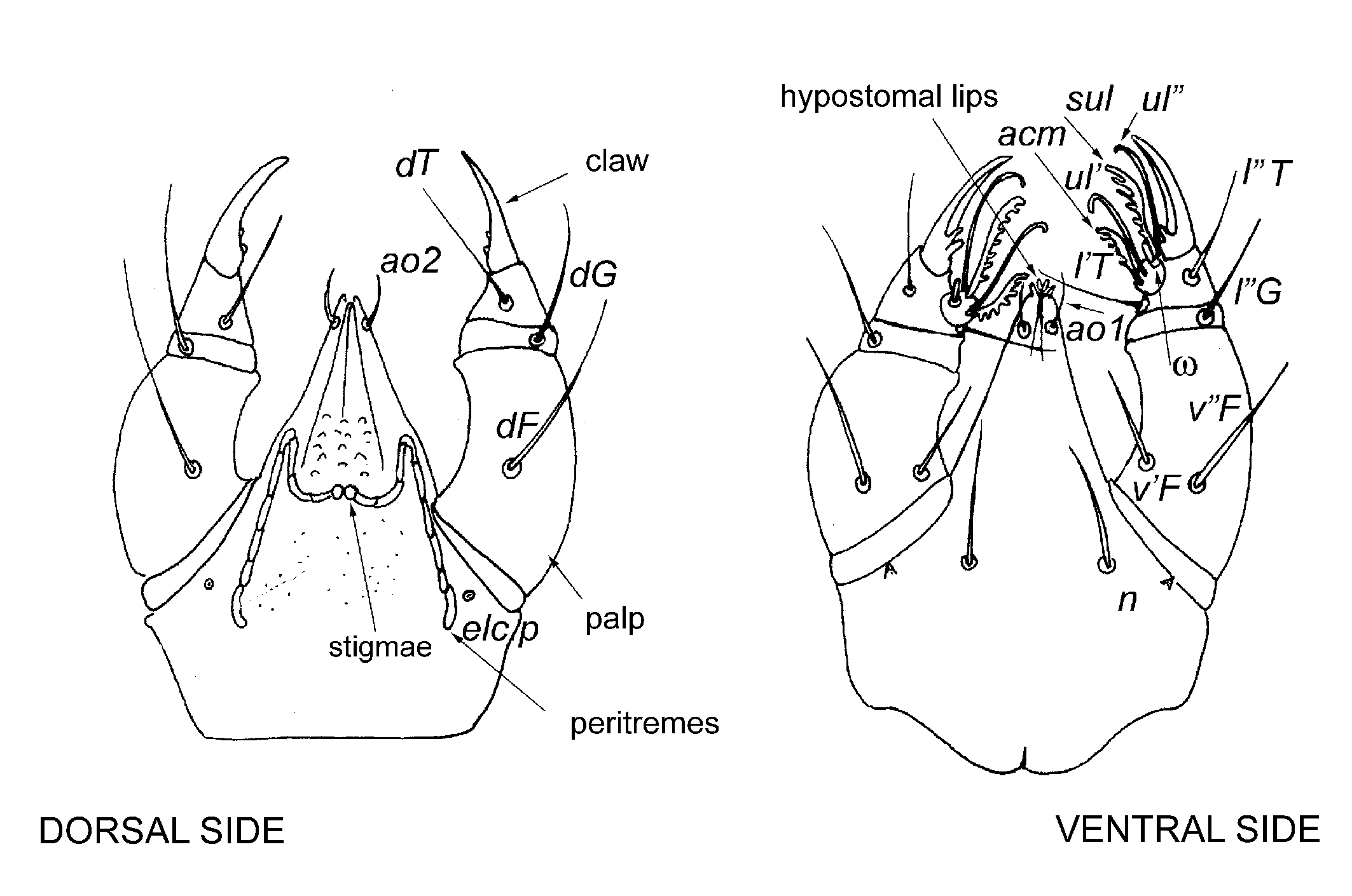

Figs.1-3. General morphology of Cheyletidae: 1. Setation of legs I-IV and tarsus I; 2. Gnathosoma; 3. Chaetotaxy of the body. (Click on miniatures for HD-image).
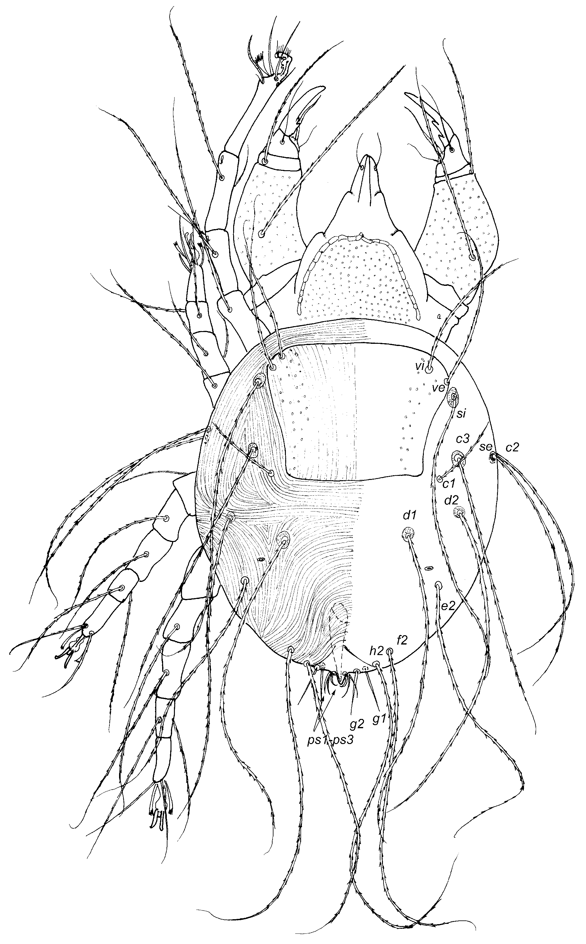
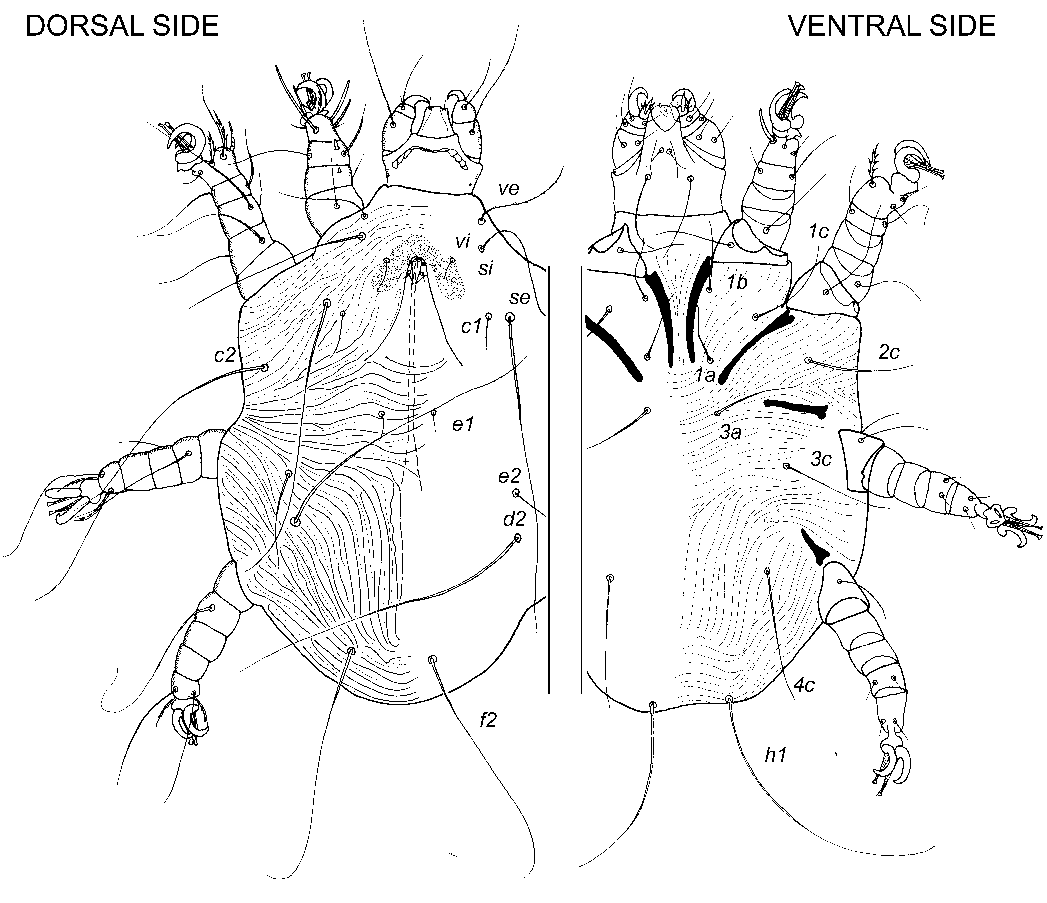


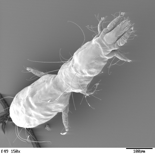
Figs. 1-5. Representatives of Cheyletidae: 1. Cheyletid male (Metacheyletoides); 2. Ornitocheyletiini male; 3. Ornitocheyletiini female; 4, Metacheyletiini female; 5, Cheletosomatiini female. (Click on miniatures for HD-image).
FAMILY SYRINGOPHILIDAE LAVOIPIERRE, 1953
Type genus: Syringophilus Heller, 1880
The external morphology of the family Syringophilidae was detailed described in two monographs: Kethley 1970 and Skoracki 2011.
 Description. Gnathosoma moderately developed. Subcapitulum deeply inserted into idiosoma, bearing setae elcp, n, ao1 and ao2; subcapitular apodeme distinctly developed. Peritremes situated on rostral part of stylophore, distinctly segmented, generally M-shaped. Hypostomal lobes fused with stylophore. Hypostomal lips present. In many taxa, female hypostome bearing 1 pair of hyaline protuberances. Palps linear, consisting of 4 segments (tibia and tarsus fused). Palpal tibiotarsus without claw. Palpal setation: femur with setae d, v’,v”, genu - d, l”, tibiotarsus - d, lT, l’, eupathidia acm, sul, ul’, ul”, and solenidion w. Idiosoma strongly elongated. Bases of trochanters inserted on ventral side of idiosoma. Eyes absent. Propodonotal shield present. Hysteronotal and pygidial shields present or absent. Ventral side of idiosoma generally devoid of shields. Opisthosoma distinctly developed. Female anal and genital-ovipore openings situated ventrally close to posterior end of body or terminally, adnate, and covered by pair of folds. In males, aedeagus tube-like and pointed apically, genital and anal openings fused (or anal opening absent) and situated dorsally. Distance between coxal fields II and III subequal or about twice as long as idiosomal width. Idiosomal cupules absent. Idiosomal setation (maximum set): scx, vi, ve, si, se, c1, c2, d1, d2, e2, f1, f2, h1, h2, ps1, ps2, ag1, ag2, ag3, g1, g2. Setae scx covered dorsally by extending lateral margins of propodonotum. In males, 2-3 pairs of aggenital setae, 2 pairs of genital setae, and 2 pairs of pseudoanal setae present. In some taxa, aggenital setae neotrichous. Legs slender, consisting of 5 articulated segments, and their tarsi having weakly developed pretarsi bearing 2 smooth lateral claws and empodium with tenet hairs. Maximum set of leg setae (paired setae are in parentheses): tarsi I-IV — I, ft, (tc),(a), (p),(u), vs, w1; II, (tc), (p),(u), vs, w1; III, (tc), (p),(u), IV, (tc), (p),(u); tibiae I-IV - I, d, (l), v,j; II, d, (l), v, j; III, d, (l); IV, d, (l); genua I-IV – I, d, l’, s; II, d, l’; III and IV l’, femora I-IV –I and II, d, v, III and IV d; trochanters I-IV, v; III; coxae I-IV – I, 1a, 1b, 1c; II, 2c; III, 3a, 3b, 3c; IV, 4b, 4c. Solenidion ω1II situated dorsally. Larva, proto- and tritonymphs present; deutonymph absent. Male moulting from tritonymph.

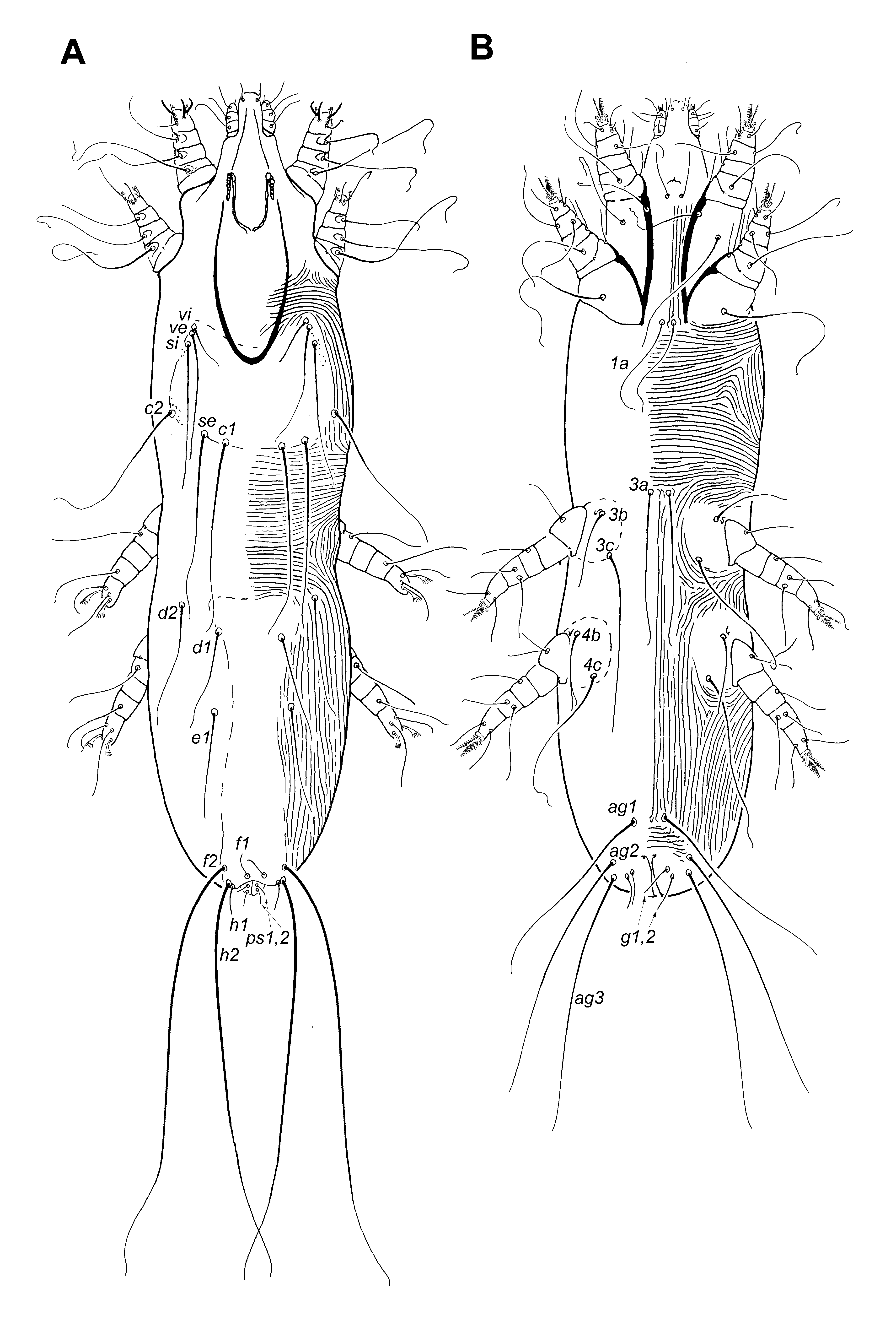
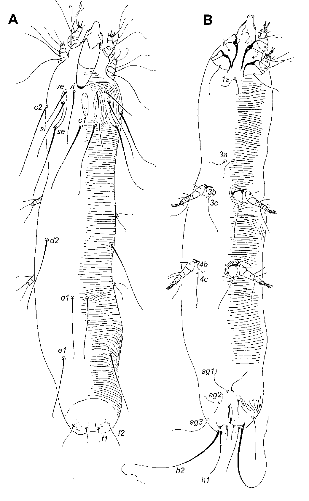
Figs.1-4. General morphology of Syringophilidae: 1. Setation of legs I-IV; 2. Syringophilinae female; 3. Syringophilinae male; 4. Picobinae female (non-physogastric form); 5. Picobiinae female (physogastric form). (Click on miniatures for HD-image).
FAMILY HARPIRHYNCHIDAE VOLGIN, 1957
The external morphology of the family Harpirhynchidae, including Ophioptinae, is described in several works (Fain 1964, 1994, 1995; Lombert and Moss 1983; Bochkov et al. 1999; Bochkov 2008; Skoracki et al. 2012). We follow here to Grandjean (1939, 1944) for the leg and idiosomal chaetotaxy. This family is probably polyphyletic and consists of three subfamilies, Harpirhynchinae (bird parasites) and Harpypalpinae (bird parasites) – Ophioptinae (snake parasites). For this reason separate diagnoses for the subfamilies Harpirhynchinae and Harpypalpinae associated with birds are provided.
Subfamily Harpirhynchinae Volgin, 1957
Type-genus: Harpirhynchus Megnin, 1877
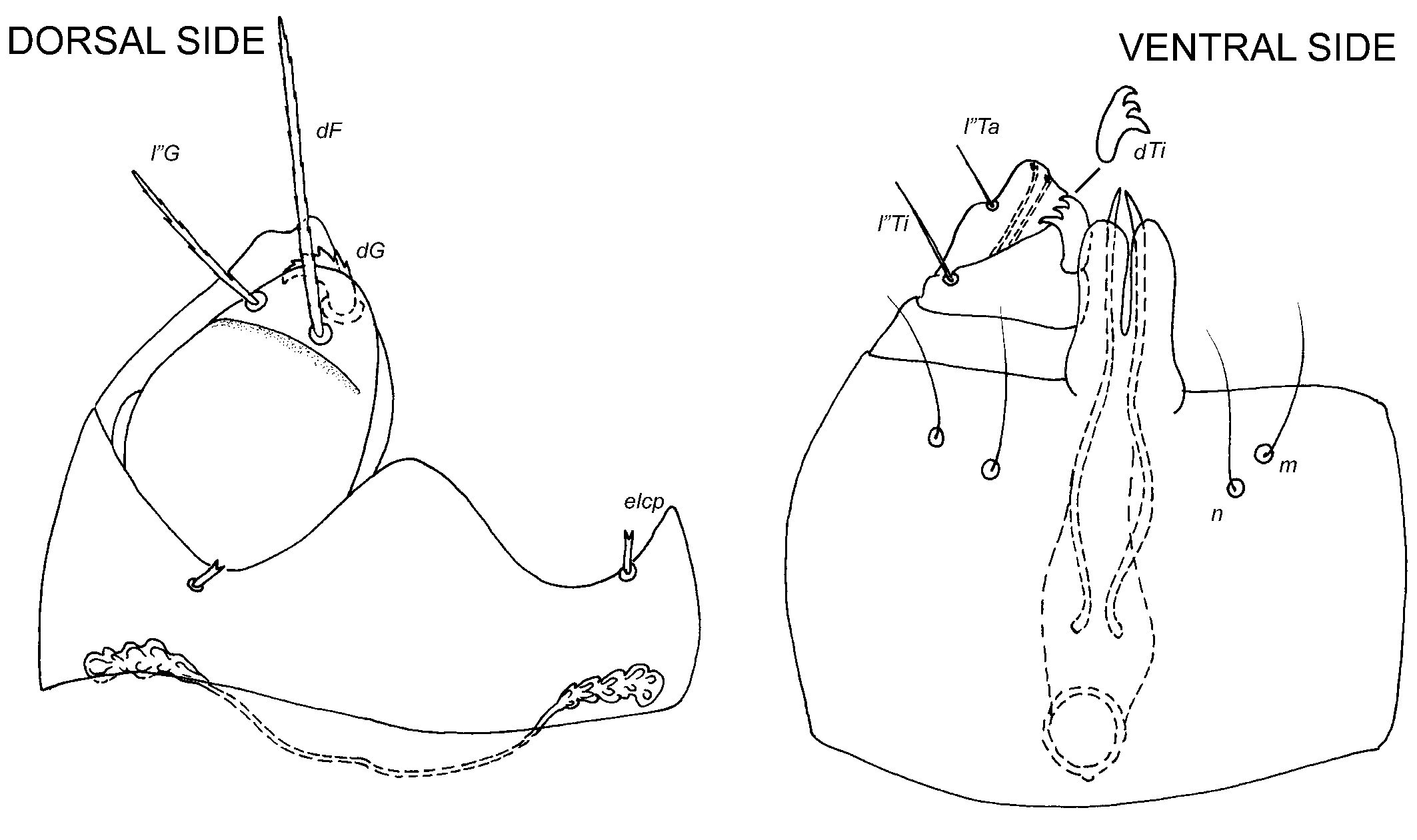
 Description. Gnathosoma moderately developed. Subcapitulum bearing setae elcp, n, and m. Adoral setae absent. Peritremes situated at base of stylophore, segmented only in distal parts, straight. Hypostomal lobes not fused with stylophore. Hypostomal lips rudimentary. Palps linear, consisting of 2 articulated segments – trochanter-femur-genu (proximal segment) and tibia-tarsus (distal segment). Palpal tibia replaced on ventral surface of trochanter-femur-genu and bearing distinct paraxial thickened seta with 2 tips (dTi) and antiaxial filiform seta (l”Ti). Palpal tarsus strongly reduced, membranous, fused with palpal tibia and bearing filiform anitaxial seta l”Ti. Proximal segment usually bearing 3 pairs of thickened setae (palpalae): dF, dG, and l”G, situated apically. In some genera setae l”G absent, and in Metharpyrhynchus mossi 1 additional palpalae present. Filiform seta v”F usually present in median part of this segment. In males, idiosoma flattened dorso-ventrally and rhomboid in outline; opisthosoma weakly developed. In females of harpirhynchines, idiosoma flattened dorso-ventrally and rhomboid in outline, sacciform (in mites living at the quill bases) or dome-shaped (in intracutaneous mites). Sacciform idiosoma could having at its posterior end “wheelbarrow” bearing lied eggs. Opisthosoma always weakly developed. In some females, idiosoma bearing various lateral and terminal lobes. Bases of trochanters inserted ventro-laterally. Eyes absent. Dorsal shield present, distinctly developed. Ventral side of idiosoma devoid of shields. Idiosomal cuticle is distinctly striated, at least, in dorsal part. In some females, striations accompanied by granules, scales, or tubercles. In females, genital and anal openings fused and situated ventrally, modified into vulvar slit, this slit could very long (up to half of idiosomal length). Unpaired pocket-like vulvar fold situated immediately before anterior end of vulvar slit. Vulvar slit could flanking by 1 pair of internal vulvar sclerites. Aedeagus well-developed, its position and length highly variable. Full set of idiosomal setae: scx, vi, ve, si, se, c2, h1, h2 (series h present only in females), 1a, 1c, and 3a; g1-3 in males. Setae scx rounded apically. Legs. Legs I and II consist of 5 articulated segments, although in females of some genera, legs I and II reduced, their segments fused and, for example, in some Metharpyrhynchus spp., they 1-segmented (fused tibia and tarsus). Tarsi I and II bearing pretarsus with paired claws and ciliated empodium. In some females with reduced anterior legs, claws and empodium strongly reduced and empodium without tenet hairs. Ambulacrum without protrusions. In both sexes, posterior legs III and IV strongly modified consisting of maximum 2 segments. Distal leg segment without ambulacrum. In females of some genera, posterior legs completely reduced or represented only by setae. Maximum set of leg I and II setae: tarsus I, tc’, tc”, p’, p”, a’, a”, u’, u”, ω1; tarsus II tc’, tc”, p”, a’, a”, u’, u”, ω1, tibia I, d, l, l”, v’, v”; tibia II, d, l, l”, v’, v”;genu I, d, l’, v’, v”; genu II, d, l’, v’, v”; femur I, d, v (could be duplicated or even triplicated); femur II, d, v (could be dupli- or triplicated); trochanter I, v, trochanter II, v. Solenidion ω1II situated dorsally. Preapical segment of legs IIII and IV bearin 1-2 setae or without setae; apical segment bearing several setae. Immature stages bearing at least anterior legs. Larva, proto- and tritonymphs present; deutonymph absent. Male moulting from tritonymph.


Figs.1-4. General morphology of Harpirhynchinae: 1. Setation of legs I-II and tarsus I; 2. Gnathosoma; 3. Harpirhinchinae male; 4. Harpirhynchinae female. (Click on miniatures for HD-image).
Subfamily Harpypalpinae Fain, 1972
Type-genus: Harpypalpus Dubinin, 1957
Description. Gnathosoma moderately developed. Subcapitulum bearing setae elcp, n, and m. Adoral setae absent. Peritremes situated at base of stylophore, segmented only in distal parts, straight. Hypostomal lobes not fused with stylophore. Hypostomal lips rudimentary. Palps linear, consisting of 2 articulated segments – trochanter-femur-genu (proximal segment) and tibia-tarsus (distal segment). Palpal tibia replaced on ventral surface of trochanter-femur-genu and bearing distinct paraxial thickened seta with 3 tips (dTi) and antiaxial filiform seta (l”Ti). Palpal tarsus strongly reduced, membranous, fused with palpal tibia and bearing filiform anitaxial seta l”Ti. Proximal segment bearing 3 pairs of thickened setae (palpalae): dF, dG, and l”G, situated apically. Seta v”F absent. Idiosoma flattened dorso-ventrally and rhomboid in outline; opisthosoma weakly developed. Bases of trochanters inserted ventro-laterally. Eyes absent. Dorsal shield present, distinctly developed. Ventral side of idiosoma devoid of shields. Idiosomal cuticle is distinctly striated in dorsal part. Coxal fields I having anteriorly directed membrane and coxal fields I–IV each with posterior membranous fold. Vulvar opening (fused anal and oviporal-genital openings) small and rounded, situated in posterior third of idiosoma ventrally. In females, it rounded by an-O or U-shaped opisthosomal membrane. In males, such U-shaped mamebrane also present. In males, the genital opening is situated dorsally. Aedeagus well-developed, and located in posterior third of idiosoma. Full set of idiosomal setae: scx, vi, ve, si, se, d1, e1, e2, f1, f2, h1, h2, ps1-3, (ag1 in females), g1 (g2 in males), 1a, 1c, 2c, 3a, 3c, and 4c. Setae scx split apically. Legs. All legs of consisting of 5 articulated segments. Ambulacra with cup-like projection, paired claws and ciliated empodium. Maximum set of leg I and II setae: tarsus I, tc’, tc”, p”, a’, a”, u’, u”, ω1; tarsus II tc’, tc”, p”, a’, a”, u’, u”, ω1, tibia I, d, l, l”, v’, v”; tibia II, d, l, l”, v’, v”;genu I, d, l’, l”, v’, v”; genu II, d, l’, l”, v’, v”; femur I, d, v (duplicated); femur II, v (duplicated); trochanter I, v, trochanter II, v. Solenidion ω1II situated dorsally. Maximum set of setae on legs III and IV: tarsus III, tc’, tc”, a’, a”, u’, u”; tarsus IV, tc’, tc”, a’, a”, u’, u”; tibia III, d, v’, v”; tibia IV, d, v’, v”; genua III and IV without setae; femur III, v; femur IV, v; trochanter III, l’, v, trochanter IV, l’, v. Immature stages without legs. Larva, proto- and tritonymphs present; deutonymph absent. Male moulting from tritonymph.
 .
. 

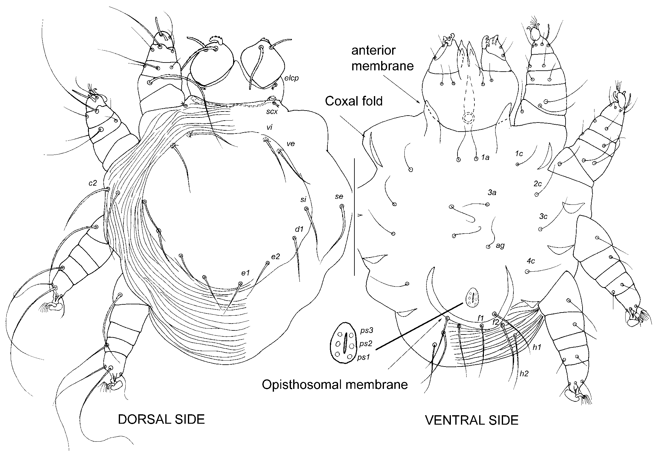
Figs.1-4. General morphology of Harpypalpinae: 1. Setation of legs I-IV and tarsus I; 2. Gnathosoma; 3. Harpirhinchinae male; 4. Harpirhynchinae female. (Click on miniatures for HD-image).
FAMILY CLOACARIDAE CAMIN, MOSS, OLIVER AND SINGER, 1967
Type genus Cloacarus Camin, Moss, Oliver and Singer, 1967
A single representative of the subfamily Pneumophaginae was recorded from lungs of Bubo virginianus
Description. Gnathosoma positioned ventrally, strongly reduced, oriented perpendicular to idiosomal axis, retracted into idiosoma, and surrounded ventrally by idiosomal wall. In Cloacarinae, remnants of posterior subcapitulum represented by distinctly sclerotized subcapitular ring, opening anteriorly and located immediately under dorsal surface of idiosoma. Pair of strongly sclerotized, fang-like chelicerae present ventrally. In Pneumophaginae, female gnathosoma devoid of posterior remnants of subcapitulum (subcapitular ring) and internal median apodemes. In males, gnathosoma completely absent. Idiosoma elliptical and slightly flattened dorso-ventrally. Propodonotal shield present and occupying most of propodonotal surface, distinctly ornamented in Cloacarinae, and weakly sclerotized and devoid of ornamentation in Pneumophaginae. Opisthosoma distinctly shorter than metapodosoma; opisthosomal striations absent. In females, anal and genital openings fused to each other forming single opening situated ventrally, terminally or dorsally. In females of some genera, lateral walls of genito-anal opening elongated and modified into 1 pair of short, finger-like projections. In Chelonacarus, genito-anal opening covered ventrally by pair of triangular sclerotised folds. In males, aedeagus present and genital opening situated dorsally on anterior part of body. Coxae completely fused with ventral surface of idiosoma; only their anterior margins represented by distinct coxal apodemes. Coxal apodemes strongly elongated, longer than half of idiosomal width. In adults, coxal apodemes I fused forming sternum. In Cloacarinae, sternum with internal median keel, at least in anterior half. Keel continues anteriorly as median apodeme with bifurcate apex. Apices fused anteriorly with subcapitular remnants and associated with sclerites, interpreted here as sigmoid pieces (= capitular apophyses). In females ofPneumophagus, anterior parts of coxal apodemes I fused with anterior part of subcapitular remnants. Proximal parts of coxal apodemes I-IV with 2 condyles at trochanteral articulation. Dorsal condyle shifted onto ventral side of idiosoma due to coxal flattening in these mites. Ventral condyles distinctly smaller than dorsal condyles. Idiosomal setae strongly reduced in size and number. In Cloacarinae, female propodonotum with 1-2 pairs of alveoli. Opisthosoma of cloacarine females bearing 2-4 pairs of setae. Setation of male propodonotum similar to that of females; opisthosoma with 1 pair of terminal, fleshy setae. In females of Pneumophagus propodonotum with 1 pair of alveoli, opisthosoma with 3 pairs of short spine-like setae situated dorsally, terminally and ventrally. Pair of alveoli flanking genitor-anal opening dorsally. In males, only 1 pair of short spine-like setae present, situated dorsally on opisthosoma. Legs I-IV with 3 articulated segments, very short; rudimentary segment inserted proximal to apical segment. Two basal leg segments devoid of setae. Trochanters largest leg segments. Posterior margins of all trochanters with large triangular projection articulated with respective coxal apodemes. In most cloacarids reduced tibiae with 1 spine-like seta, but devoid of setae in Pneumophagus, except for 1 seta on tibia I. Tarsi broadly rounded in cloacarines; in Pneumophagus tarsi II-IV flattened laterally. Claws absent on all tarsi. In Cloacarinae tarsi I and III each with 8 setae (in Caminacarus chrysemys Pence and Casto, 1975, tarsi III with 9 setae), tarsi II – 9 setae, and tarsi IV – 7 setae. Short globose solenidion on tarsi I-III situated ventro-terminally. In Pneumophagus; tarsi I-IV bearing 5-9-12-12 setae, respectively. Solenidion absent on all tarsi. Larva unknown. Only 1 nymphal stage known.


Figs. 1 and 2. General morphology of Cloacaridae: 1. Pneumophagus bubonis, female and male; 2. Gnathosoma and leg II. (Click on miniatures for HD-image).
FAMILY EREYNETIDAE OUDEMANS, 1931
Type genus Ereynetes Berlese, 1883
The taxonomic content of the family Ereynetidae was reviewed by Fain (1957). He included the
family Speleognathidae erected by Womersley
(1936) to the Ereynetidae as a subfamily together
with the newly created Lawrencarinae.
In 1985, Fain divided the subfamily Speleognathinae into five tribes: Boydaiini, Trispeleognathini, Speleognathini, Speleochirini, and Paraspeleognathopsini. Among them, representatives
of the two first tribes are associated with birds.
Description. Chelicerae bases fused to each other and with subcapitulum forming gnathosomal capsule. Movable cheliceral digit short, linear and stylet-like. Palps linear non-raptorial, composed of 4 segments (trochanter, femuro-genu, tiba, tarsus) in Ereynetinae and reduced to 1-3 segments in Lawrencarinae and Speleognathinae. Claw-like structures correspond to strong ventral setae (v) and observed only in Ereynetinae. Palpal setation, femur-genu with 2 pairs of dorsal setae dF and dG (in Ereynetinae, absent in other subfamilies), tibiae with 2 dorsal setae t’ and t” in Ereynetinae, 1 or without setae in other subfamilies, tarsus with setae d, l’, l”, pζ, v, in Ereynetinae, reduced to 2-3 setae (d, l’, l”) in Lawrencarinae and Speleognathinae, tarsal solenidion ω present in Ereynetinae and present or absent in other subfamilies. Hypostomal apex with 1-2 pairs of subcapitular setae (sc1, sc2). Postcheliceral stigmata obscure. Idiosoma weakly sclerotized, sac-like. Reticulate ornamentation of prodorsum present or absent. Eye lenses present or absent. Propodonotal and hysteronotal shields present in some ereynetines and absent in most of speleognathines and Lawrencarinae. Ventral side of idiosoma without plates. Distance between coxal fields II and III distinctly shorter than idiosomal width. Idiosomal setation (maximum in Ereynetinae): vi, ve, si (= anterior sensilla), se, c1, c2, d1, e1, f1, f2 (= posterior sensilla), h1. Pair of posterior sensilla usually present in free-living species and absent in most parasitic species. Cupules im and ia present or absent. Genital opening situated behind legs IV, longitudinal. Genital region with setae: ag1-5, ps1-2, g1-5. Neotrichy of setae ps observed only in Lawrencarus eweri (Lawrencarinae). In males of Ereynetinae 1-3 pairs of eugenital setae (gi) present, in Lawrencarinae and Speleognathinae 1 pair of setae gi present or absent. Genital papillae 2 pairs, well developed in Ereynetinae, or reduced in Lawrencarinae and Speleognathinae. Legs long and massive, covered by reticulate ornamentation. Coxal fields of legs I-IV distinctly bordered and well sclerotized. Coxae setation (maximum in Ereynetinae) I-IV: I 1a, 1b, 1c; II 2c; III 3a, 3b, 3c, 3d; IV 4a, 4b, 4c. Setae 3d and 4c absent in Lawrencarinae and Speleognathinae. Trochanters I-IV – I-III l’; IV without setae. Femora I-IV – I with 4-7 setae; II (d), (v); III v, d, l; IV (d), (v). Femora I, IV divided on basi- and telofemur in Ereynetinae, not in Lawrencarinae and Speleognathinae. Genua I-IV – I (d) (l); II (d), (l); III d, (l); IV d, (l). Tibiae I-IV – I d, (l), (v); II d, (v), III d, (v); IV d (v). Tibiae I with internal sensory complex (ereynetal organ) where solenidion of tibia I (φI) recessed into inverted sac-like structure with narrow duct that opens near distal margin of segment. Tarsi I-IV - I holotrichous with 12 setae, solenidion ω present or absent; II with 9 setae, III and IV with 8 setae. Tarsi I-IV with a pair of large claws and with setiform median empodium. In development 5 postembryonic instars: larva, protonymph, deutonymph, tritonymph and adult. All nymphs mobile in ontogenesis of Ereynetinae and Lawrencarinae, while in Speleognathinae all nymphs are calyptostatic.

Fig. 1. General morphology of Ereynetidae. (Click on miniatures for HD-image).